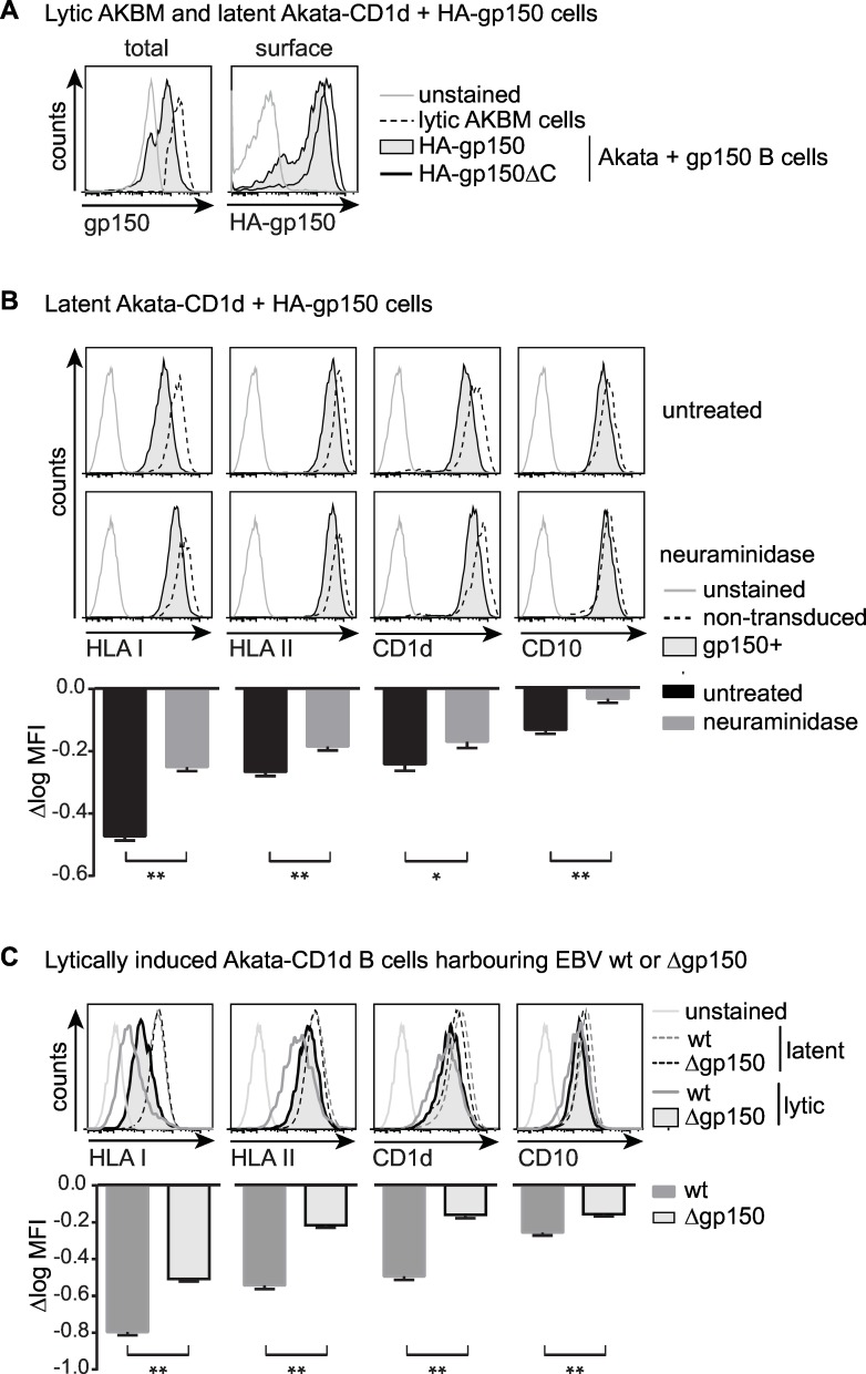
Fig 7. Glycan shielding of surface Ag-presenting molecules by gp150 occurs in human B cells and is reversed during productive EBV infection when gp150 is deleted.
A-B) Latent AkataΔgp150 cells were lentivirally transduced to co-express either HA-gp150 or HA-gp150ΔC and GFP (from EF1a and PGK promoters, respectively) and were puromycin-selected to obtain pure populations of gp150+ cells. B cells already grow in suspension and gp150-positive B cells were maintained in culture for several weeks, indicating that gp150 expression was not toxic to the cells. A) Flow cytometry was used to assess total (intracellular staining with anti-gp150 Ab on permeabilized cells) and surface (anti-HA Ab on non-permeabilized cells) levels of EBVgp150. Expression levels of gp150 were compared to lytically induced AKBM cells (20 hours anti-human IgG treatment, rat CD2GFP+ cells). B) Akata+gp150 and non-transduced control cells were left untreated or were treated with neuraminidase (1U/μl, 60 min, 37°C). Surface levels of Ag-presenting molecules and cellular CD10 as a control were compared to gp150- non-transduced cells. One representative experiment of three is depicted. C) Viral reactivation was induced in Akata wt and Δgp150 B cells by overnight culture with anti-human IgG and EBV-producing cells were identified by staining for the late viral protein gp350. Surface levels of Ag-presenting molecules and CD10 on lytic (gp350+) and latent (gp350-) cells are depicted in overlay histograms. One representative experiment of at least four is depicted. Statistical analysis was performed for B and C) as described for Fig 6. * p<0.05, ** p<0.01.

