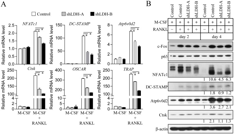
Fig 4. Altered osteoclastogenic signaling during osteoclast differentiation of LDH-A or LDH-B-deficient cells.
Osteoclast precursors were infected with LDH-A, LDH-B, or pLKO.1-puro empty control virus particles. LDH-A or LDH-B knockdown cells were cultured with M-CSF (30 ng/ml) and RANKL (100 ng/ml) for 3 days (A) or the indicated days (B). (A) The mRNA expression levels of osteoclastogenic genes including NFATc1, DC-STAMP, Atp6v0d2, cathepsin K (Ctsk), OSCAR, and TRAP were analyzed using quantitative real-time PCR. Data are mean ± SD (n = 3). *P < 0.01. (B) Total cell lysates were subjected to immunoblot analysis with specific antibodies to c-Fos, p65, NFATc1, DC-STAMP, Atp6v0d2, cathepsin K (Ctsk), and β-actin (loading control). Band intensities were represented as a fold difference. Gel images are representative of three independent experiments.

