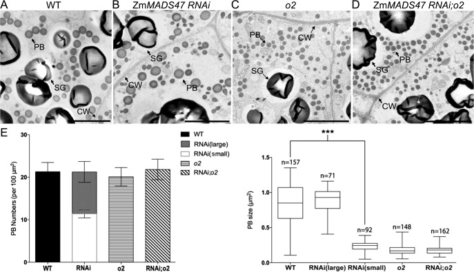
Fig 4. Transmission electron microscopy of wild type, ZmMADS47 RNAi, o2, and ZmMADS47 RNAi; o2 starchy endosperm.
A-D. Protein bodies in the fourth starchy endosperm cell layer at 18DAP stage were observed by transmission electron microscopy. Each genotype is labeled above the corresponding TEM image. PB: protein body; CW: cell wall; SG, starch granule. Bars = 1μm. E. Quantitative comparison of PB number and size in the fourth and fifth cell layer from aleurone between wild type, ZmMADS47 RNAi, o2, and ZmMADS47 RNAi;o2 endosperm. Error bars represent SD (***P < 0.001, Student’s t test).

