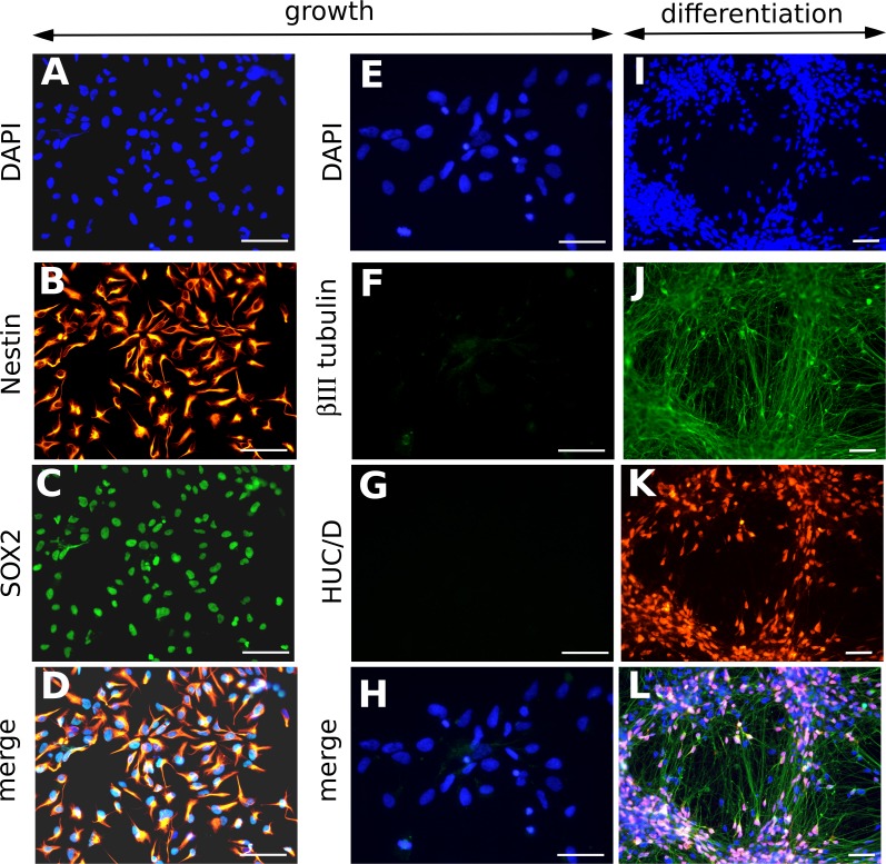
Fig 1. Characterization of neural stem cells from human ES cells (NSCs).
Representative immunofluorescence analyses of NSCs cultured in growth medium (A-H) or in differentiation medium, 8 days after the onset of differentiation (I-L), using DAPI staining (A, E, I), or antibodies specific to Nestin (B), SOX2 (C), βIII tubulin (F, J) and HUC/D (G, K). Merged pictures are shown (D, H, L). In differentiation medium, neurons positive for βIII tubulin (J) and HUC/D (K) went alongside to undifferentiated NSCs, which nuclei appeared blue in the merged picture (L). Scale bar: 50 μm.

