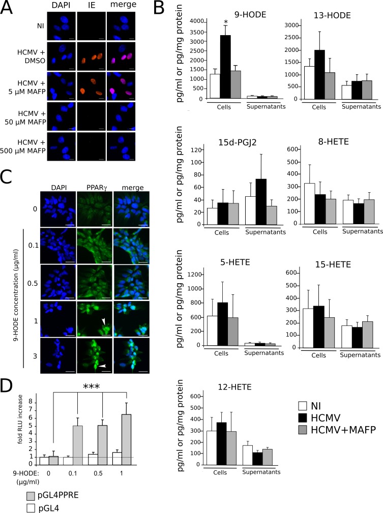
Fig 6. Increased production of PPARγ agonist 9-HODE in infected NSCs.
(A) Immunofluorescence analysis of IE expression, showing that treatment of the HCMV inoculum by the PLA2 inhibitor MAFP impairs IE expression. NI: uninfected cells in medium containing 50 nM MAFP. (B) LC-MS/MS screening of PUFA-derived lipids produced in NSCs infected by live (HCMV) or MAFP-treated (HCMV+MAFP) HCMV, or in uninfected NSCs (NI), showing significant increase in 9-HODE levels in infected NSCs (top left). Amounts in supernatants are expressed in pg/ml, amounts in cell lysates are expressed in pg/mg protein. Data represent means ± CI of a minimum of 5 independent experiments, each being performed in triplicate. *: p<0.05. (C) Immunofluorescence analysis of PPARγ expression in uninfected NSCs showing increased PPARγ levels and nuclear translocation (arrowhead). (D) Luciferase assay showing increased PPARγ activity in NSCs stimulated by 9-HODE. HCMV strain was AD169. Data represent means ± CI of 2 independent experiments, each being performed in triplicate. ***: p<0.005.

