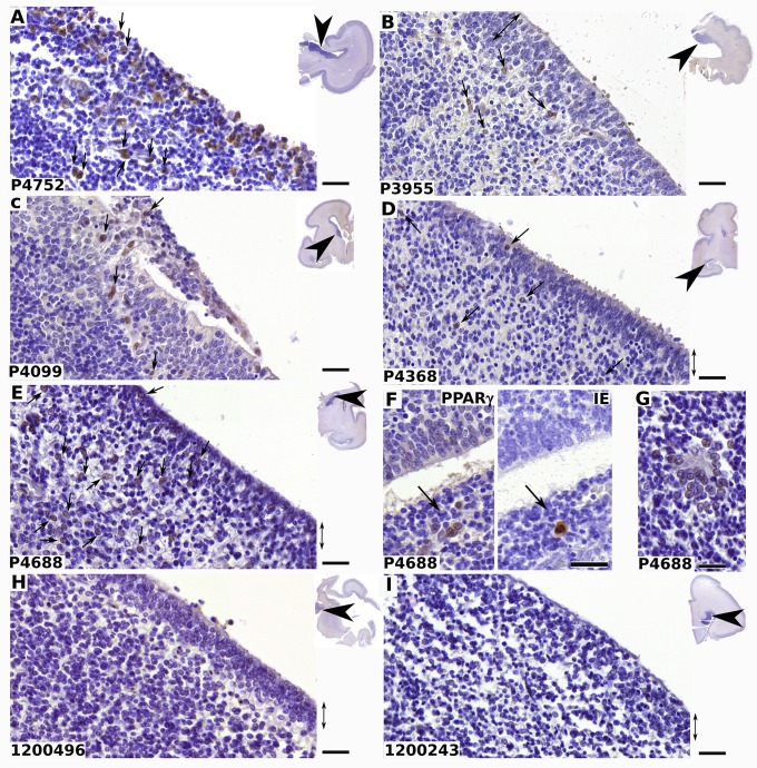
Fig 9. Nuclear PPARγ expression in germinative zone of HMV-infected human fetal brains.
Shown are representative results of immunohistological staining of brain sections from fetuses infected by HCMV (A-G) or from controls (H, I) using antibodies against PPARγ (A-E; F, left; G-I) or IE (F, right). The reference number of each donor is indicated at the bottom left of each panel. Clinical details are summarized in Table 1. PPARγ positive cells (arrows) are detected in the germinative, periventricular, areas and in ependyma (double arrow) in cases, but not in controls. Insets show the localization of the optical field within the brain sections (arrowheads). Note the nuclear localization of PPARγ (A-G), the presence of PPARγ positive cell islets surrounding one IE positive cell in two fields from serial sections (F) and clusters of PPARγ immunoreactive cells around lesional tissue (G). Scale bar: 50 μm.

