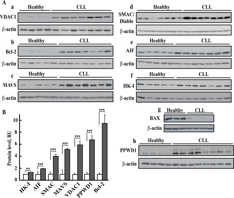
Fig 3. Over-expression of Bcl-2, VDAC, AIF, MAVS, SMAC/Diablo and PPWD1 in PBMCs from CLL patients.
Immunoblot analysis of cell lysates of PBMCs derived from CLL patients (P) and healthy donors (H) probed with antibodies directed against VDAC1 [Aa, n = 28 (P), 20 (H)], Bcl-2 [Ab, n = 28 (P), 20 (H)], MAVS [Ac, n = 28 (P), 19 (H)], SMAC/Diablo [Ad, n = 21 (P), 15 (H)], AIF [Ae, n = 17 (P), 16 (H)], HK-I [Af, n = 28 (P), 20 (H)], BAX [Ag, n = 6 (P), 6 (H)], PPWD1 [Ah, n = 16(P), 10(H)] or β-actin. Representative immunoblots (A) and quantitative analysis (mean ± SEM) (B) of protein levels of healthy donors (white) and CLL patients (grey) of these and other samples are presented. For each sample, three independent immunoblots were performed. A difference between healthy and CLL groups was considered statistically significant when P < 0.001 (***) or P < 0.01 (**), as determined by the Mann-Whitney test.

