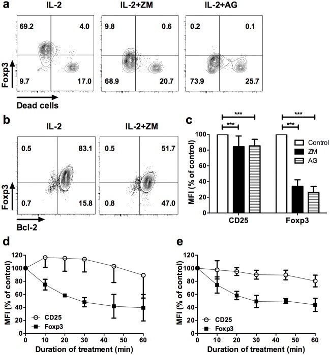
Fig 2. Reduced Foxp3 by JAK/STAT inhibitors is independent of cell death.
(a) CD4+GFP+ T cells sorted from Foxp3-GFP transgenic mice were stimulated with anti-CD3/CD28 beads for 8 days and treated with vehicle and IL-2 (IL-2), or IL-2 supplemented with ZM (IL-2+ZM) or AG (IL-2+AG) JAK inhibitors for two hours. Cells were then stained with a dye allowing exclusion of dead cells by flow cytometry compatible with Foxp3 intracellular staining. Profiles are representative of 6 and 7 independent experiments for AG and ZM inhibitors, respectively. (b) Foxp3 and Bcl-2 expression in gated CD4+CD25+ cells from expanded CD4+GFP+ cells after 2 hour treatment with vehicle control and IL-2 (IL-2) or ZM in presence of IL-2 (IL-2+ZM). Similar results were observed in 3 independent experiments. (c) Median fluorescence intensity (MFI) of CD25 and Foxp3 in dead cell–excluded subset, represented as percentages of the MFI relative to Foxp3 staining in vehicle control condition (control) after two hours treatment with the indicated inhibitors. The results are compiled from 6 (AG) and 7 (ZM) independent experiments. (d) Freshly isolated or (e) anti-CD3/CD28 activated CD4+GFP+ cells from Foxp3-eGFP reporter mice were treated for the indicated times with either ZM or AG inhibitors. MFI of CD25 and Foxp3 are relative to the MFI of vehicle control conditions. These data are compiled from 3 independent experiments. Statistical significance was tested using Student t-test (***p<0.001, **p<0.01, *p<0.05).

