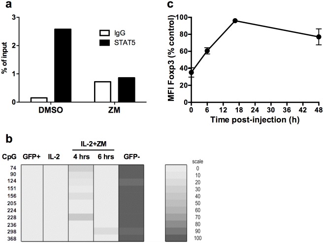
Fig 4. Loss of Foxp3 is not due to remethylation of the CNS2 and is reversible.
(a) STAT5 binding on a putative binding site in the TSDR enhancer of the foxp3 gene was assessed by chromatin immunoprecipitation with control IgG or anti-STAT-5 antibodies. Quantification of binding was performed as described in Methods section. These data are from one representative experiment out of two. (b) Methylation status of the Foxp3 TSDR was determined by bisulphite sequencing. Expanded CD4+GFP+ T cells were treated with IL-2 and ZM (IL-2+ZM) for 4 (4 hrs) or 6 (6 hrs) hours. Freshly purified CD4+GFP+ (GFP+), CD4+GFP- (GFP-), or untreated expanded cells (IL-2) were used as controls. These data are representative of two independent experiments. Each line indicate the position in the Foxp3 locus (CpG), according to Floess et al. [11]. A shaded scale of grey illustrates the frequency of each methylated CpG. (C) For in vivo analysis, expanded CD4+GFP+ cells treated or not with AG for two hours were injected into congenic C57BL/6 mice. Animals were euthanized at the indicated times after transfer. The MFI of Foxp3 was normalized to vehicle-treated control cells.

