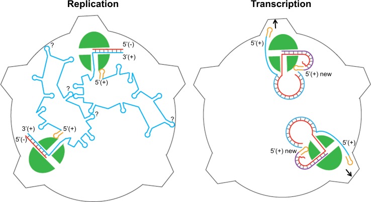
Fig 8. Proposed model for hPBV RdRP replication (left) and transcription (right) inside the viral capsid.
The hPBV RdRP molecules are displayed in green. The blue and red lines correspond to the (+) and (-)strand respectively with the nascent (+)strand RNA represented in purple (right). The 5’-terminal stem loop structure is displayed in yellow. How exactly the two ssRNA molecules interact with each other and also with the viral CP during assembly and replication is not yet clear, as indicated by the questions marks in the figure on the left. The parental (+)strand RNA is separated from the template RNA during transcription and directed towards a pore in the viral capsid (right).

