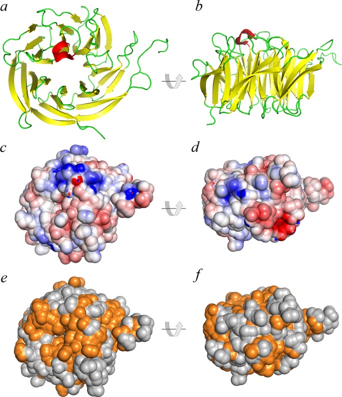
FIGURE 2:

Structural features of WDR92. (a, b) Ribbon diagrams of H. sapiens WDR92 from the 1.95-Å crystal structure (3I2N; Xu and Min, 2011); the β−strands are indicated in yellow and the single short α-helix is in red. The two views are related by a 90° rotation. (c, d) The same orientations of WDR92, shown with the van der Waal molecular surface displayed and painted based on a full Poisson–Boltzmann electrostatics calculation; negatively charged regions are in red and positively charged segments in blue. (e, f) Residues on the WDR92 molecular surface are colored orange to illustrate those regions that are completely conserved between the H. sapiens, S. mediterranea, and C. reinhardtii orthologues; orientation is the same as before.
