FIGURE 5:

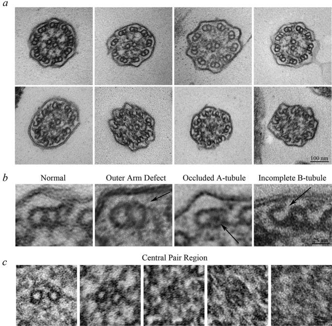
Smed-wdr92(RNAi) planaria have defective ciliary architecture. (a) Transmission electron micrographs of transverse sections through cilia of Smed-wdr92(RNAi) animals. Although many cilia appear morphologically normal (top left), others exhibit a variety of structural defects, including missing outer dynein arms, occlusion of the A-tubule lumen, accumulation of electron-dense material within the central region of the axoneme, where it obscures (or replaces) the central pair microtubules, and the incomplete closure of B-tubules at the outermost A/B-tubule junction. Bar, 100 nm. (b) Magnified views of individual microtubule doublets illustrating the variety of structural defects observed. Bar, 25 nm. (c) Magnified views of the central pair region of the axonemal lumen. Although most axonemes contained a morphologically normal central complex (left), others lacked the central pair and/or contained amorphous material that either obscured or replaced it. In these cases, the assembly status of the radial spokes was also unclear. Bar, 25 nm.
