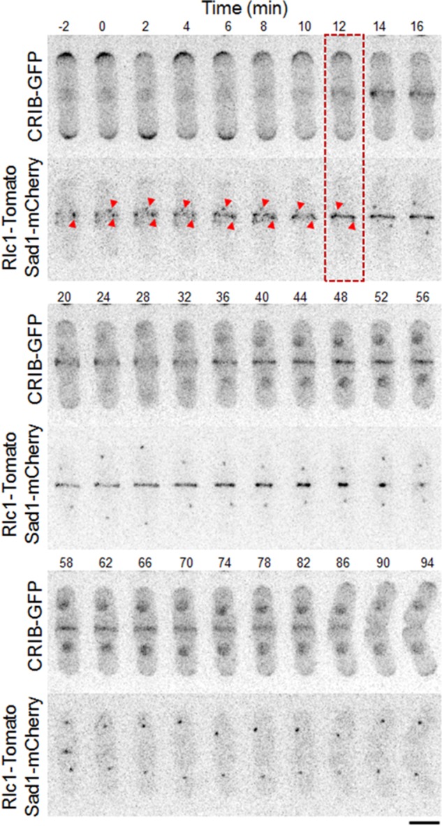
FIGURE 1:

Cdc42 is activated at the site of cell division during cytokinesis. Time-series images showing the appearance and duration of the CRIB-3xGFP signal at the site of cell division. SPB separation by Sad1-mCherry and the cytokinetic ring protein by Rlc1-Tomato (bottom). Red arrowheads show initial stages of SPB marker position. Red box depicts onset of Cdc42 activation. Bar, 5 μm. Time is in minutes.
