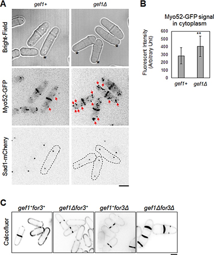
FIGURE 6:
Gef1 promotes type V myosin Myo52 localization to the cell division site. (A) Type V myosin Myo52-GFP distribution in gef1+ and gef1Δ cells. Cells with comparable distance of SPB marker Sad1-mCherry were selected. Bright-field images ensured nonseptating cells were selected. Red arrows mark cytoplasmic Myo52-GFP patches. Asterisks mark cells in the ring phase of cytokinesis. (B) Quantification of Myo52-GFP signal in the cytoplasm in gef1+ and gef1Δ cells as described; 22 cells, **p = 0.0015. Error bars, SD. (C) Calcofluor staining of gef1+for3+, gef1Δ, for3Δ, and gef1Δfor3Δ cells grown at 35°C. Bars, 5 μm.

