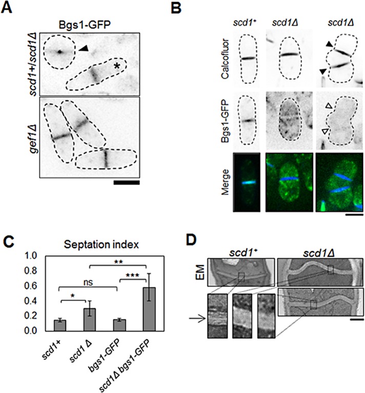
FIGURE 8:
Scd1 is required for normal septum formation. (A) Cells expressing Bgs1-GFP in scd1+ and scd1Δ cells analyzed in the same field. scd1+ cells are depicted by asterisks, and scd1Δ cells are depicted by arrowheads. Bottom, Bgs1-GFP–expressing gef1Δ cells shown for comparison. Bar, 5 μm. (B) Calcofluor staining of scd1+ and scd1Δ cells expressing Bgs1-GFP. scd1Δ cells with multiple septa (arrowheads). Bar, 5 μm. (C) Quantification of septation index in cells as indicated; >223 cells. *p < 0.05, **p < 0.01, ***p < 0.001; ns, not significant. Error bars, SD. (D) Transmission electron microscopy of septum in scd1+ and scd1Δ cells. Sections of septum (black box) are zoomed at 4× to show primary septum defects. Bar, 500 nm.

