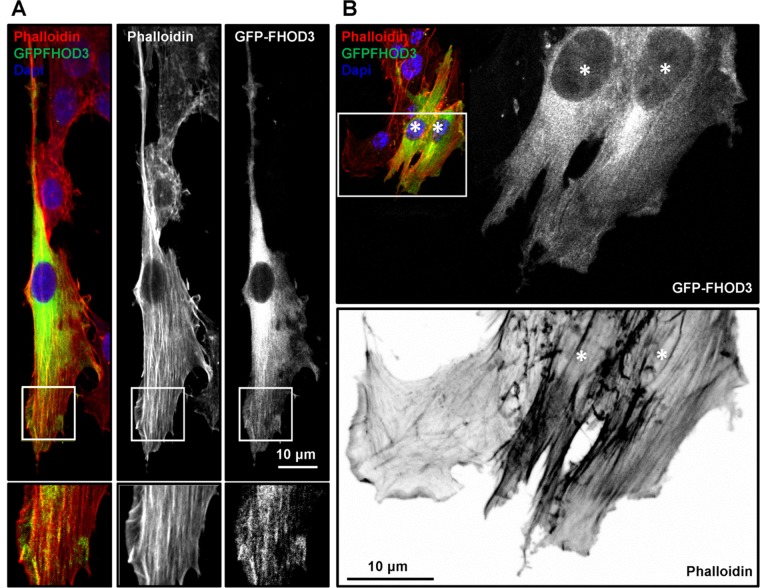
FIGURE 8:
GFP-FHOD3 localizes on parallel actin filaments found in leading processes. C6 glioma cells were transfected with GFP-FHOD3 and seeded on 20-μm laminin micropatterned lines (A) and 2D substrates (B). Cells were fixed and stained with phalloidin and 4′,6-diamidino-2-phenylindole and imaged using a confocal microscope. (B) GFP-FHOD3–overexpressing cells (asterisks) among untransfected cells display excessive amounts of longitudinal actin bundles. Scale bars, 10 μm.

