FIGURE 1:
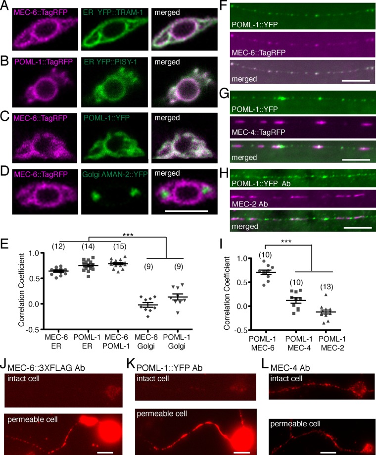
TRN expression of MEC-6 and POML-1. Confocal sections of TRN cell bodies of (A) MEC-6::TagRFP and the ER marker YFP::TRAM-1, (B) POML-1::TagRFP and the ER marker YFP::PISY-1, (C) MEC-6::TagRFP and POML-1::YFP, and (D) MEC-6::TagRFP and the Golgi marker AMAN-2::YFP and their correlation coefficient (E). Scale bars, 5 μm (A–D, F–H, J–L). The number of examined TRNs is given in parentheses (E, I). Symbols for significance here and in all subsequent figures are described in Materials and Methods. Neurite expression of (F) POML-1::YFP and MEC-6::TagRFP, (G) POML-1::YFP and MEC-4::TagRFP, and (H) POML-1::YFP and MEC-2 and their correlation coefficient (I). Anti-GFP and anti-MEC-2 antibodies (Ab) were used to label the proteins in (H). (J) MEC-6::3XFLAG expression as detected by an anti-FLAG antibody in intact (top) and permeabilized (bottom) cultured TRNs. (K) POML-1::YFP expression as detected by an anti-GFP antibody in intact (top) and permeabilized (bottom) TRNs in culture. The faint immunofluorescence in intact cells (J, K) was not specific because it was often observed in cells that did not express MEC-6::3XFLAG or POML-1::YFP. Images in J and K are representative of 40 cells examined in two independent experiments. (L) MEC-4 expression detected with an anti-MEC-4 antibody that recognizes the extracellular domain in intact (top) and permeabilized (bottom) cultured TRNs. Images are representative of 20 cells examined in two independent experiments.

