FIGURE 7:
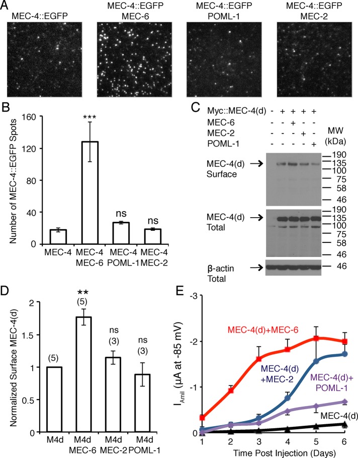
Effect of MEC-6 and POML-1 on MEC-4 surface expression in Xenopus oocytes. (A) Images and (B) quantification of MEC-4::EGFP fluorescent spots on the oocyte surface visualized by TIRF imaging (19–29 patches from 14–16 cells from two different batches) 2 d after cRNA injection. Values are compared with the expression of MEC-4::EGFP alone using the Mann–Whitney test. The field dimensions are 13 μm × 13 μm. (C) Western blot of Myc::MEC-4(d) on the surface of oocytes as detected by biotinylation (top) and the expression of Myc::MEC-4(d) in total lysate of oocytes (middle) at 2 d after cRNA injection. β-Actin detected in total lysate was used as an input control (bottom). Molecular weights (kilodaltons) of the protein markers are indicated on the right. (D) Quantification of changes in surface Myc::MEC-4(d) detected by biotinylation at 2 d after cRNA injection (the number of independent experiments is given in parentheses). All data are normalized and compared with Myc::MEC-4(d) expression alone by the one-sample t test. MEC-6, MEC-2, and POML-1 did not affect Myc::MEC-4(d) levels in total lysates at 2 d after injection (MEC-6, 1.0 ± 0.1; MEC-2, 1.0 ± 0.1; POML-1, 0.9 ± 0.1; four or five independent experiments, normalized and compared with the expression of Myc::MEC-4(d) alone; not significant by one-sample t test). The normalized amount of total Myc::MEC-4(d) differed by no more than 25% in any of the experiments. (E) The amiloride-sensitive MEC-4(d) current at −85 mV (12–18 oocytes [2 d after cRNA injection] or 6–12 oocytes [other times] of three batches) on its own and in the presence of MEC-2, MEC-6, and POML-1 at various times after cRNA injection. p < 0.001 for Iamil at −85 mV between oocytes 2 d after injected with MEC-4(d) and MEC-6 vs. MEC-4(d) alone, MEC-4(d) and MEC-2, or MEC-4(d) and POML-1; no statistically significance was found between oocytes 2 d after injection with MEC-4(d) alone vs. MEC-4(d) and MEC-2, or MEC-4(d) and POML-1. p < 0.001 for Iamil at −85 mV between oocytes 1 d after injected with MEC-4(d) and MEC-6 vs. MEC-4(d) alone; p < 0.01 between MEC-4(d) and MEC-6 vs. MEC-4(d) and MEC-2, or MEC-4(d) and POML-1. One-way ANOVA with Tukey post hoc.

