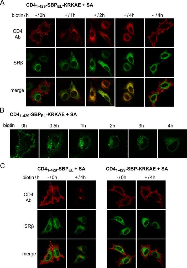
FIGURE 8:
Controlled transport of plasma membrane reporter to the ER. Cells were cotransfected with CD41–429-SBP–based constructs and SA (1:1). After 24 h, cells were incubated with anti-CD4 antibodies in the cold, washed, and incubated at 37°C for the indicated times with or without biotin. (A, C) Cells cotransfected with CFP-SRβ were fixed and stained with CY3-conjugated secondary antibody. (B) Uptake of Alexa 488–conjugated anti-CD4 antibody was followed by live imaging.

