FIGURE 6:
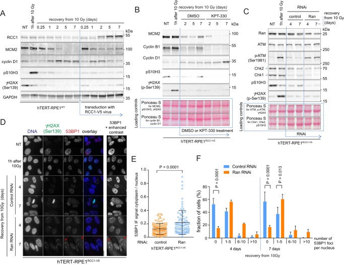
RCC1-induced activation of NCT promotes the completion of the DNA repair and cell cycle reentry. (A–C) Immunoblots showing the changes of the cell cycle and DDR markers in cells exposed to different treatments after the γ-irradiation (10 Gy). (A) The γ-irradiated hTERT-RPE1WT cells were transduced with control or RCC1-V5 expressing lentiviruses. (B) The γ-irradiated hTERT-RPE1RCC1-V5 cells were treated with dimethyl sulfoxide (control) or 500 nM KPT-330. (C) Control or Ran-directed RNAi was applied after the γ-irradiation of the hTERT-RPE1RCC1-V5 cells. (D) Micrographs of 53BP1 and γH2AX IF staining in the hTERT-RPE1RCC1-V5 treated as in C. Inset with the enhanced contrast shows the accumulation of the cytoplasmic 53BP1 signal in the Ran RNAi-treated cells (arrows). (E) Scatter plot of the cytoplasmic/nuclear ratios of the 53BP1 IF signal in the control or Ran RNAi-treated cells at 7 d of recovery from the γ-irradiation. Individual cell data; means ± SD; t test, representative of two experiments. (F) Fractions of hTERT-RPE1WT and hTERT-RPE1RCC1-V5 cells recovering from γ-irradiation that contained the indicated numbers of 53BP1 foci per nucleus. Means ± SD from two independent experiments; two-way ANOVA with Bonferroni’s posttest.

