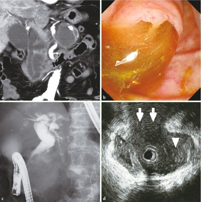
Fig. 1 a.

Computed tomography (CT) image showing multiple cystic lesions with septa in the pancreatic head and body, and a suspected fistula connecting to the common bile duct (CBD). b Endoscopic appearance of the duodenal papilla: dilated pancreatic and bile ducts with mucin extrusion (“pig-nose” appearance). c Endoscopic cholangiogram revealing a dilated biliary tree in which the distal and mid CBD have an extensive filling defect. d Intraductal ultrasonography (IDUS) showing a pancreatobiliary fistula. The white arrows show the pancreatobiliary fistula. The white triangle shows the CBD.
