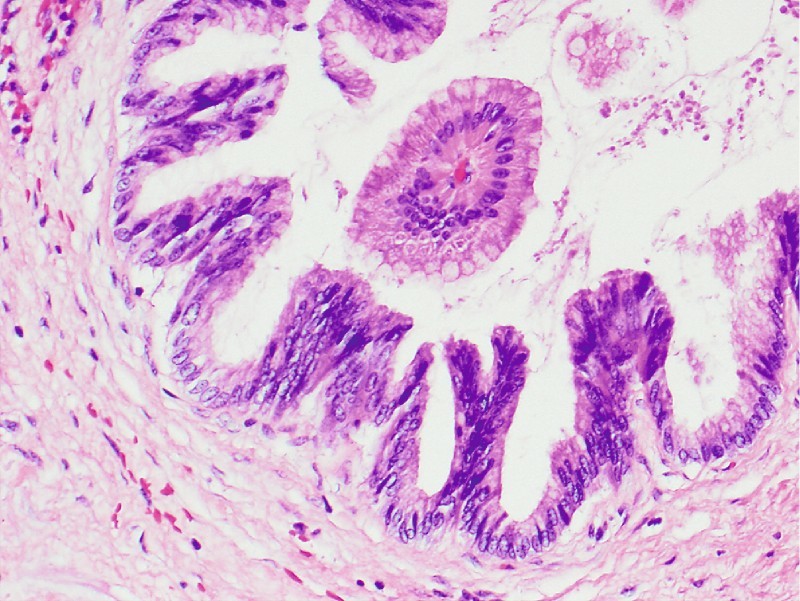
Fig. 5.

Surgical specimen, case 1 ( × 20). Gastric-type intraductal papillary mucinous neoplasm with intermediate grade dysplasia (cellular crowding, nuclear stratification, loss of polarity). Note the fibrovascular papillary core surrounded by well-oriented mucinous epithelium without dysplasia.
