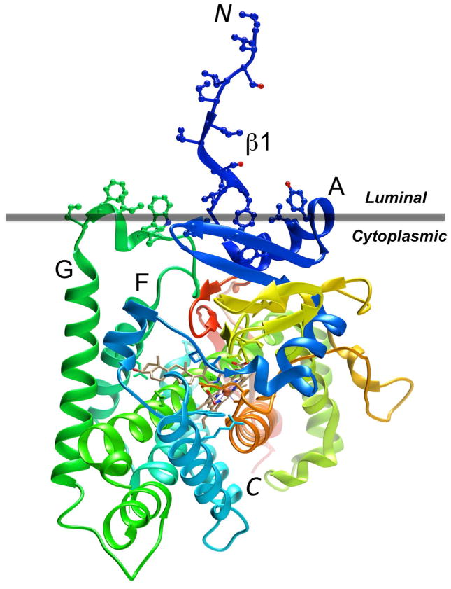
Figure 1.
Tertiary structure diagram of human P450 17A1 (PBD ID code 3RUK; http://www.rcsb.org) with the ribbon colored in the rainbow spectrum, from N-terminus (blue) to C-terminus (red). Amino acid side chains in hydrophobic regions of the protein associated with the membrane (horizontal gray bar) are depicted in ball and stick mode. The P450 enzyme is located on the cytoplasmic side of the bilayer (Monk et al., 2014). The image was generated with the program UCSF Chimera (Pettersen et al., 2004).

