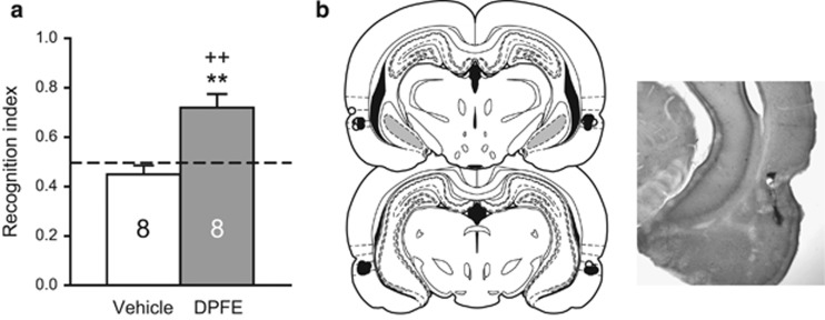
Figure 5.
Intraperirhinal DPFE restored novel object recognition in the same cohort of animals tested in the novel cue relapse test. (a) Novel object recognition testing from Experiment 3. **p<0.01 compared with vehicle; ++p<0.01 compared with a hypothetical mean of 0.50. (b) Histological verification of microinjector needle placements within perirhinal cortex from Experiment 3. The distribution of vehicle placements are indicated by white dots; black dots indicate DPFE placements. Infusions were predominantly clustered in area 35, just below the rhinal fissure. A section from a representative animal is shown at right.

