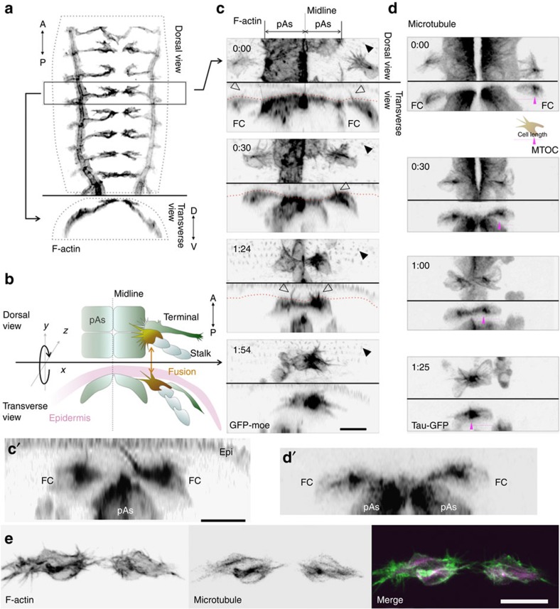
Figure 1. Dorsal-branch migration and fusion of the Drosophila trachea.
(a) A stack of confocal GFP images shows the tracheal system of a stage-15 embryo carrying btl>GFP-moe. (b) Spatial relationship of the migrating tracheal branch and the epidermis; FCs, terminal cells and stalk cells of the dorsal branch and peripheral amnioserosa (pAs) are indicated. (c) Time course of dorsal-branch migration and fusion, shown by F-actin labelling with esg_FC-Gal4 and UAS-GFP-moe (FC and pAs), and with sqh-GFP-moe (epidermis). Each panel shows dorsal (top half) and transverse views (bottom half) of images at the indicated time points (top left of the image). Filled triangles indicate F-actin-rich denticles of the apical surface of the epidermis; open triangles indicate FC filopodia penetrating vertically into the epidermis and nearly reaching the apical surface. Red dotted line indicates the basal epidermal surface. (c') Enlarged view of the vertical filopodia (1:30). (d) Microtubule distribution during tracheal-branch fusion, revealed by expressing the tau-GFP marker in FCs and pAs by the esg_FC-Gal4 driver. Magenta triangles indicate MTOCs. (d') Enlargement of d, 0:30 time point. (e) Double labelling of microtubules (GFP-tau) and F-actin (GFP-moe). Scale bar, 10 μm.

