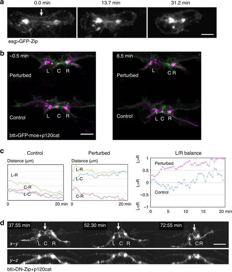
Figure 3. FC contraction.
(a) GFP-myosin heavy chain formed a longitudinal track connecting two adherens junctions in an FC. (b) Laser perturbation of FC contraction: the left-side cell (asterisk) in a new FC pair was illuminated by infrared laser at the time of contact. (c) The location of each cell-adhesion site (L, C and R) was tracked and the distance between sites was plotted. Laser perturbation caused significant deviations in L/R balance values (N=3, a representative case is shown). (d) Fusion was inhibited by a dominant-negative form of myosin. White arrow indicates the FC contact site. Scale bar, 10 μm.

