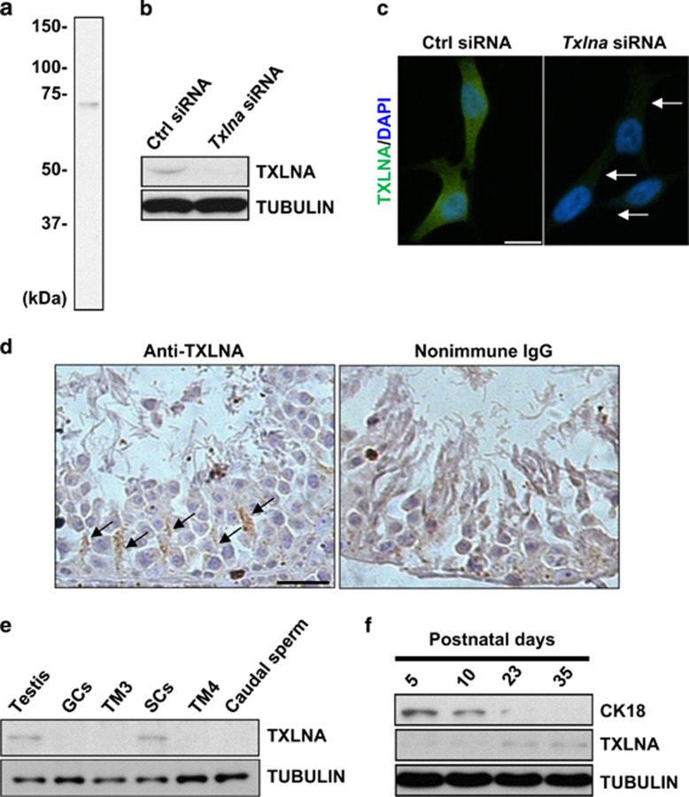
Figure 1.
TXLNA is predominantly expressed in functionally mature Sertoli cells (SCs). (a) Immunoblotting analysis demonstrated a single band of TXLNA protein in the testicular lysates, which was absent when samples were incubated with preabsorbed primary antibody or without primary antibody. (b) The efficiency of the targeted knockdown of Txlna in 3T3 cells was confirmed by western blotting at the translational level. Tubulin served as a loading control. (c) The efficiency of the targeted knockdown of Txlna in 3T3 cells was also confirmed by immunofluorescence staining. Arrows indicate positive signals of TXLNA protein. Scale bar, 10 μm. (d) Immunohistochemical analysis in murine testes revealed a distinct cytoplasm localization of TXLNA in SCs (arrows). Replacement of the primary antibody with nonimmune IgG abolished the immunostaining, confirming the specificity of the assay. Scale bar, 25 μm. (e) Expression profile of TXLNA was evaluated in different spermatogenic cell lines and in mouse testis using western blotting. Tubulin served as a loading control. (f) Immunoblotting analysis of TXLNA and CK18 protein in mouse developing postnatal testis. Tubulin served as a loading control

