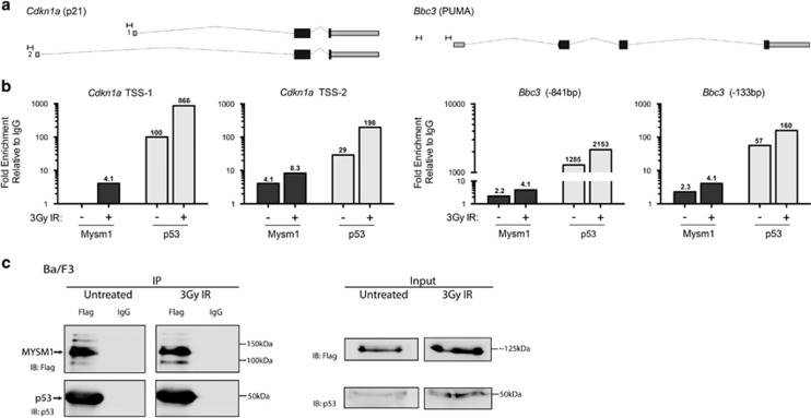
Figure 2.
MYSM1 protein can interact with p53 and colocalize to the promoters of p53-target genes. (a and b) Ba/F3 hematopoietic progenitor cells stably expressing 3xFlag-MYSM1 protein were analyzed for recruitment of MYSM1 and p53 to the promoters of p53 target genes using ChIP-qPCR at a steady state and following 3-Gy irradiation. (a) Genomic structure of Bbc3/PUMA and Cdkn1a/p21 loci according to the UCSC Mouse mm9 assembly with target primer sites and PCR products indicated (to scale). Primers were designed in the vicinity of known p53-binding sites based on ChIP-Seq data from Kenzelmann Broz D et al. (b) Fold enrichment of MYSM1 and p53 near the transcriptional start sites of Bbc3/PUMA and Cdkn1a/p21 genes in untreated and irradiated cells (3 h after 3 Gy). Relative enrichment was normalized relative to a nonspecific IgG ChIP and to a negative control genomic region (Pomc) with normalized values shown above each bar. Data shown are representative of at least three independent experiments with additional replicates in Supplementary Figures S2c and d. (c) Co-immunoprecipitation of endogenous p53 with 3xFlag-MYSM1 stably expressed in the Ba/F3 hematopoietic progenitor cell line. Cell lysates from untreated and irradiated cells (3 h after 3 Gy) were subjected to immunoprecipitation with anti-Flag and IgG control antibodies. Left panel: IP samples were blotted for Flag-MYSM1 (anti-Flag) and p53 showing specific pulldown of both proteins. Right panel: whole-cell lysates (input) were blotted for Flag-MYSM1 (anti-Flag) and p53

