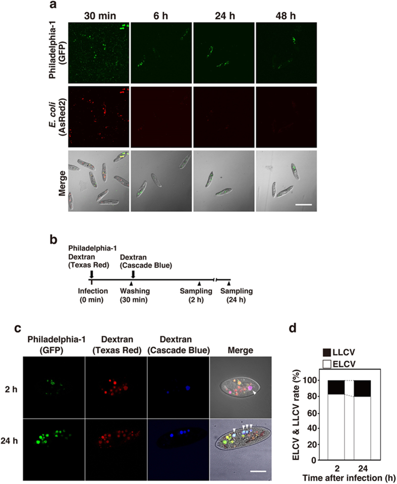
Figure 1. L. pneumophila Philadelphia-1 establishes endosymbiosis in P. caudatum RB-1.
(a) Intracellular localization of Philadelphia-1 (GFP) and E. coli (AsRed2) in RB-1 at 30 min, 6 h, 24 h, and 48 h after infection. Bacteria were simultaneously added to RB-1 at an MOI of 10000. Scale bar represents 100 μm. (b) Bacteria and each dextran were added to RB-1 according to this schedule. LCVs containing each dextran were observed by confocal laser scanning microscopy 2 h and 24 h after infection (c), and percentages of ELCV and LLCV are shown with the total of all LCVs being 100% (d). Arrowheads point to LLCV, which are LCVs positive for both Texas Red- and Cascade Blue-conjugated dextrans. Scale bar represents 30 μm.

