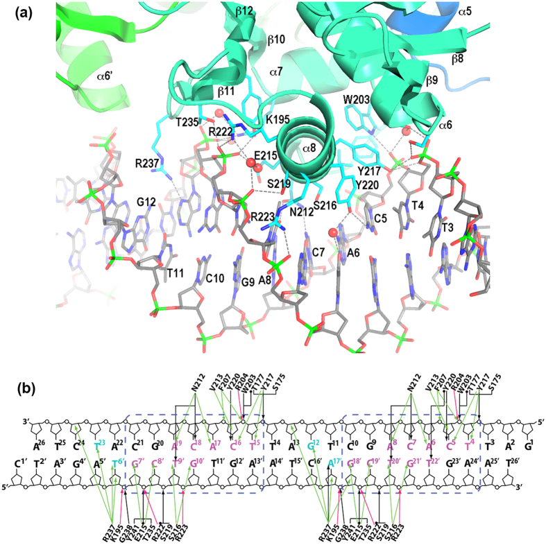
Figure 3. Details of PhoP-DNA interactions.
(a) A ribbon diagram showing the DNA-binding elements of PhoP interacting with DNA. DNA is shown in sticks. The bases in the strand containing the TCACAGC motif are labelled. The side chains of PhoP that interact with DNA are shown as sticks. The hydrogen bonds between the protein and DNA are shown as dashed lines. (b) Schematic diagram showing detailed interactions between PhoP and DNA in both subsites of the direct repeat. DNA sequences of both strands in the crystal structure are shown. The base pairs of the TCACAGC motifs are boxed in blue dashed lines. Bases in the major groove that are in contact with protein atoms are coloured magenta; those in the minor groove in contact with the Arg237 side chain are coloured cyan. The hydrogen bonds are shown as black arrows, and water-mediated hydrogen bonds are marked with a dot at the beginning of the arrows. Van der Waals contacts within 4 Å are shown as green arrows, and salt bridges are shown as red arrows.

