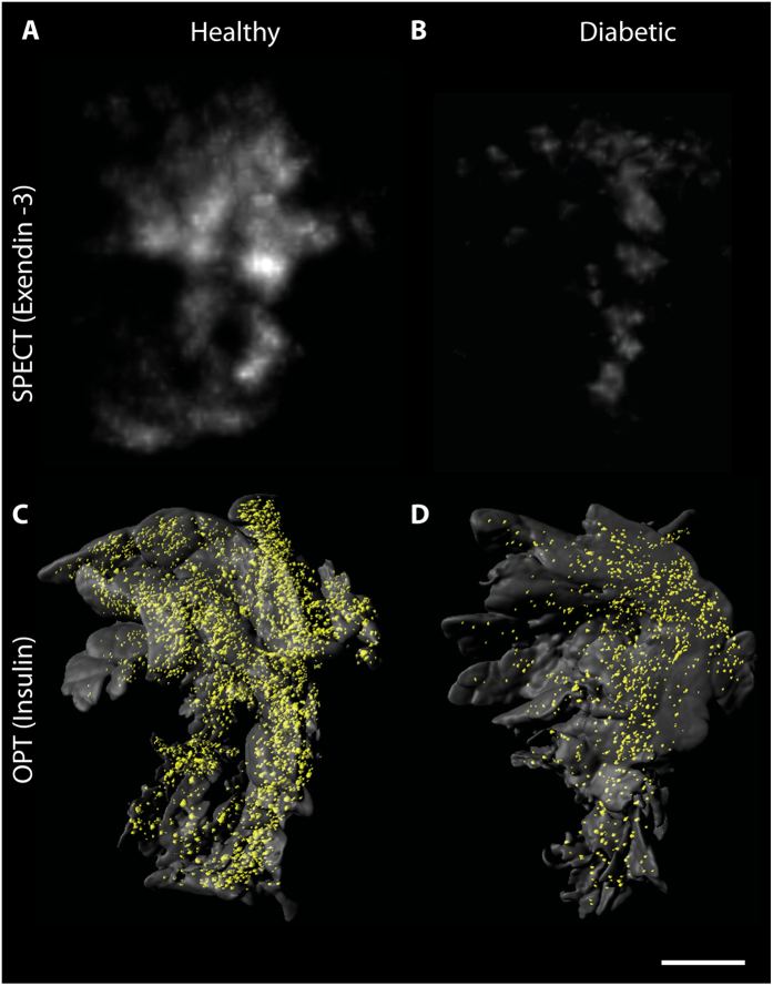
Figure 2. Multimodal imaging of pancreatic β-cells with SPECT and OPT.
(A,B) Ex vivo SPECT scans of representative splenic lobes from a healthy (A) and an alloxan treated animal (B) respectively. (C,D) OPT generated iso-surface images of the same lobes as visualized in (A,B). Alloxan-treated rats exhibit lower 111In-exendin-3 uptake and β-cell volume when compared to the control group. Scale bar represents 3 mm.

