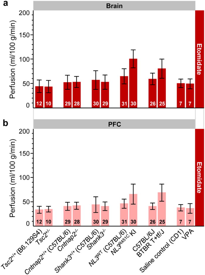
Figure 2. Comparison of basal blood perfusion in the brain and PFC of six mouse models of autism spectrum disorder under etomidate anesthesia.

Perfusion in the (a) brain and (b) PFC was assessed in spontaneously breathing Tsc2+/−, Cntnap2−/−, Shank3−/−, NL3R451C KI, BTBR T+tf/J, VPA-exposed mice, and corresponding wild-type controls. Sample sizes are provided in each bar. Error bars indicate standard deviations to visualize the spread of data for individuals. All the models were on a C57BL/6J background, except VPA (CD1) and Tsc2+/− (B6.129S4 mixed background). Wild-type littermates were used as controls, except for BTBR T+tf/J mice where age-matched C57BL/6J mice served as controls. KI, knock-in; PFC, prefrontal cortex; VPA, valproic acid; WT, wild-type.
