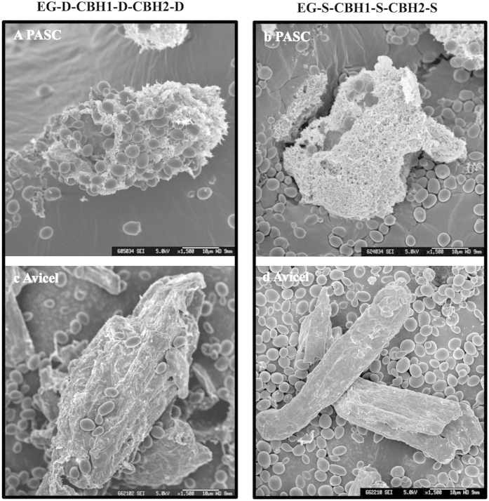
Figure 4. SEM micrographs of the interactions between cellulolytic S. cerevisiae cells and cellulosic materials.
Interactions with PASC (a,b) and with Avicel (c,d). Cellulolytic cells (30 g/L) were incubated with 1% cellulosic materials (PASC or Avicel) for 2 h, and the cellulosic materials were used for SEM imaging. Scale bars are 10 μm.

