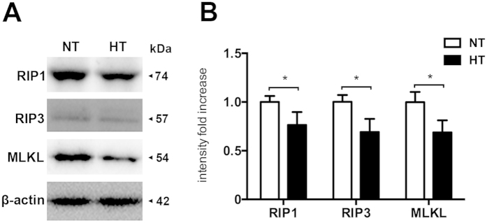
Figure 2. Hypothermia reduces necroptosis processing in the injured cortex after TBI.
Rats were sacrificed at 6 h after TBI. Total proteins were prepared from the ipsilateral cortex of TBI-Normothermia (NT) and TBI-Hypothermia (HT). (A) Immunoblot analysis of RIP1, RIP3 and MLKL in lysates of ipsilateral cortex after injury (n = 6 each). (B) Quantitative analysis of RIP1, RIP3 and MLKL after TBI. Lane-loading differences were normalized by levels of β-actin; Data are expressed as mean ± SD; Data of RIP1 are analyzed using ANOVA with Dunnett’s post-hoc test vs. SHAM. *P < 0.05.

