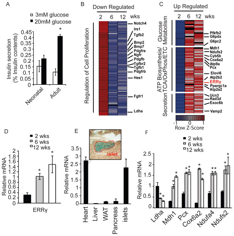
Figure 1. Islets acquire oxidative features postnatally.
(A) Glucose-stimulated insulin secretion from freshly isolated mouse neonatal (< 14 days) and adult (> 12 weeks) islets after sequential perfusion with 3mM and 20mM glucose for 30 min (reported as % insulin content, 10 islets per assay, n=6). (B–C) Heatmaps of transcriptional changes in islets during postnatal maturation. Row Z-score of down-regulated (B), and up-regulated (C) genes. (D) Relative Errγ expression in isolated islets measured by qPCR. (E) Relative Errγ expression in murine 12 wk old heart, liver, white adipose (WAT), pancreas and islets (n=4). X-gal staining indicating ERRγ expression in islets from 12 wk old ERRγ knock-in mice (top panel). (F) Relative expression of Ldha and selected mitochondrial metabolic genes during postnatal islet maturation, as measured by qPCR. (n=3). Data represent the mean ±s.e.m. *p<0.01 Student’s unpaired t-test. See also Figure S1.

