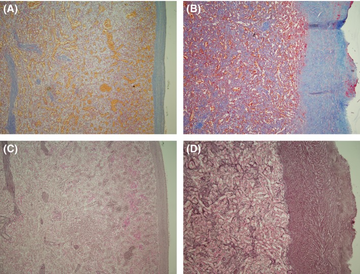
Figure 1.

Spleen histology. A & B Martius scarlet blue stain for collagen. (A) Normal spleen (control); (B) Spleen of Case 1. C & D: Reticulin stain. (C) Normal spleen (control); (D) Spleen of Case 1. Panels B & D illustrate clearly the fivefold thickening of the spleen capsule. All panels taken at low power magnification.
