
Figure 1.
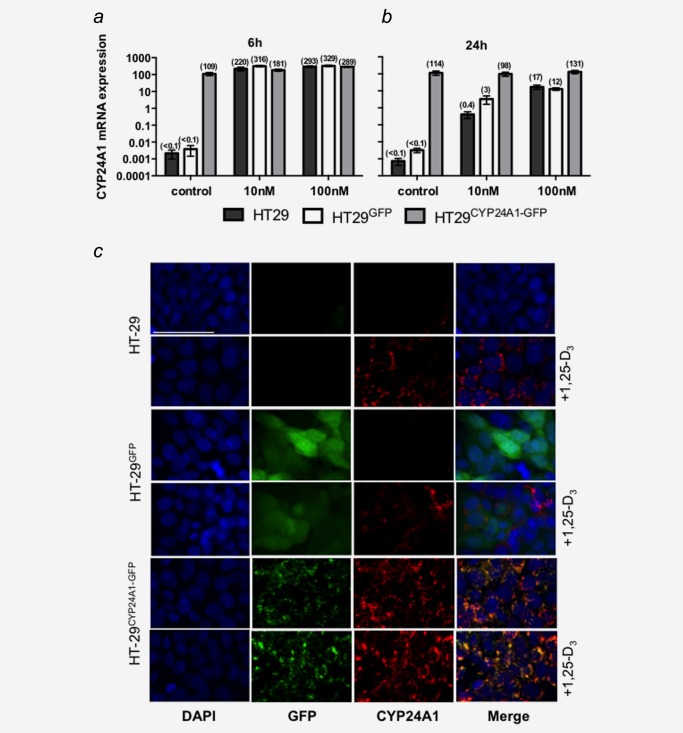
mRNA and protein expression of CYP24A1 in cell lines. The parental cell line (HT29) was stably transfected with a GFP‐tagged plasmid (HT29GFP) or a plasmid encoding for the fusion protein CYP24A1‐GFP (HT29CYP24A1‐GFP). Cells were treated for 6 h (a) or 24 h (b) with 10 nM 1,25‐D3, 100 nM 1,25‐D3 or vehicle control. mRNA levels of CYP24A1 were assessed by real time qRT‐PCR. All values were set relative to total RNA calibrator and are presented as 2−ΔΔCT. Bars represent mean ± SEM of three independent experiments. (c) Cells were stained for CYP24A1 expression on coverslips after 20 h of treatment with 100 nM 1,25‐D3, or vehicle. Vector GFP expression is detectable in HT29GFP and HT29CYP24A1‐GFP cells but is absent in the parental cell line. Basal CYP24A1 (red) protein expression is low with exception of HT29CYP24A1‐GFP cells but is inducible with 1,25‐D3. White line represents 50 μm.