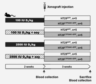
Figure 3.

Schematic presentation of the mouse model. Mice were divided into four groups receiving low (100 IU D3/kg diet) or high (2500 IU D3/kg diet) vitamin D diet ± soy (20%). After 2 weeks HT29GFP or HT29CYP24A1‐GFP cells (106 per mouse) were injected to the right flank of the mice. The experiment was terminated at the end of the 5th week from the starting of the experiment (2 weeks acclimatization to the diet plus 3 weeks of xenograft growth).
