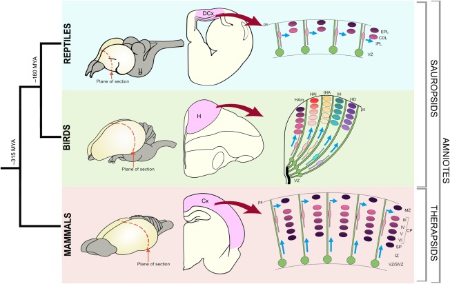
Figure 1.

Phylogenetic relationship of mammals, birds, and reptiles. Schematic representations of brains with the plane of sections (coronal) indicated (dotted red lines). Schematic representation of sections through the reptile, bird, and mammalian brains with dorsal cortex (DCx), hyperpallium (H), and cerebral cortex (Cx) shaded in pink. The generation of the cortical neurons is schematized in the right panels. In the reptilian brain the progenitors (green) divide in the ventricular zone (VZ) and the postmytotic cells produce the cortex that is comprised of internal plexiform layer (IPL), cell dense layer (CDL), and external plexiform layer (EPL). The progenitors extend from the VZ to pial surface (ps) and generate waves of neurons that are different in different sectors. The bird hyperpallium has regional variations and several radial segments can be distinguished. A more homogeneous population of neurons of the VI layers (VI–I) are generated from progenitors of the cortex. It is postulated that the neurons of different cortical layers is generated from the same progenitors in a sequential fashion.
