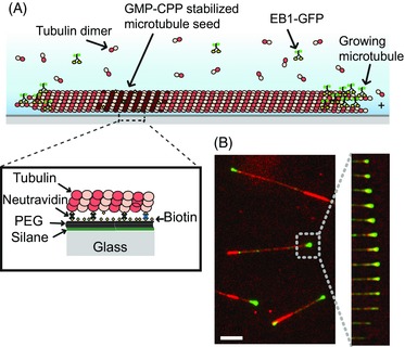
Figure 1.

Experimental dynamic microtubule assay. (A) Schematic of a dynamic microtubule attached to a glass cover slip, showing a fluorescently labelled end binding protein, such as EB1‐GFP, accumulating at the growing microtubule end. (B) Left – Example image from a TIRF microscopy movie showing EB1‐GFP (green) binding to a Cy5 labelled microtubule (red). Right – time series of successive frames of the movie showing growth and shrinkage of a microtubule end.
