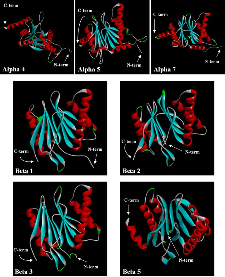
Figure 7. Schematic view of the backbone fold of seven proteasome subunits from T. bernacchii.
Alpha helix, beta-strand and turn structures are indicated with red, cyan and green colours respectively. The positions of N- and C-termini are indicated by arrows. Models were generated as described in Materials and Methods section. Images have been created with Discovery Studio software.

