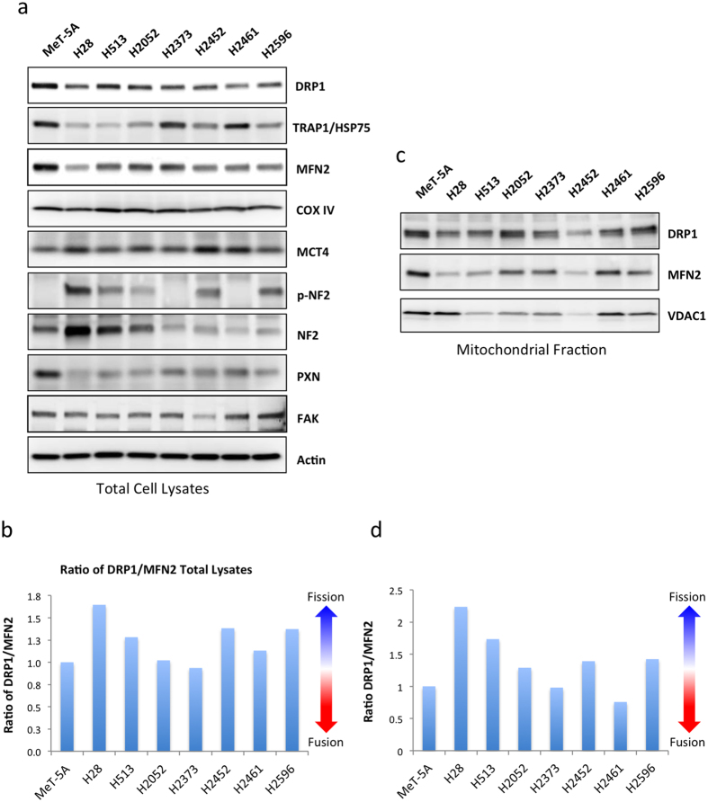
Figure 4. Altered expression of mitochondrial proteins in malignant mesothelioma cell lines.
(a) Representative immunoblots showing expression of mitochondrial and focal adhesion marker proteins in a panel of mesothelioma cell lines and a control transformed but non-tumorgenic cell line, MeT-5A. (b) Ratio of DRP1 to MFN2 expression in total cell lysates as measured via densitometry (c) Representative immunoblots showing expression of DRP1, MFN2 and VDAC in the mitochondrial fraction of mesothelioma cell lysates (d) Ratio of DRP1 to MFN2 expression in the mitochondrial fraction of mesothelioma cell lysates as measured via densitometry. 20 μg of protein was loaded per sample. Results are expressed as mean ± the standard deviation of 3 independent measurements.

