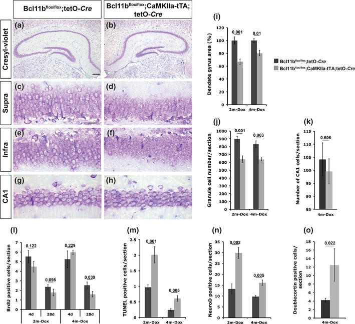
Figure 5.

Analysis of adult‐induced Bcl11b mutant mice at 2 and 4 months after doxycycline removal. (a–h) Cresyl‐violet staining of control (a,c,e,g) and Bcl11b mutant (b,d,f,h) hippocampal sections at 2 months after doxycycline removal. Scale bar, 20 µm (c); 100 µm (a). (i‐o) Quantitative analysis of the dentate gyrus area (i), granule cell number (j), CA1 cell number (k), BrdU incorporation at 4 (4d) and 28 (28d) days after the initial BrdU injection (l) as well as TUNEL (m), NeuroD (n) and Doublecortin (o) positive cells at 2 (2m‐Dox) and 4 (4m‐Dox) months after doxycycline removal. Infra, infrapyramidal blade; Supra, suprapyramidal blade; t‐test, numbers indicate P‐values; error bars, SEM; n = 3 [4m‐Dox in i,j,l,n (4d)]; n = 4 [4m‐Dox in l (28d), m; 2m‐Dox in n; mutant in o]; n = 5 (2m‐Dox in i,j); n = 8 (2m‐Dox in l; control in o).
