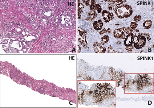
Figure 2.

IHC staining for SPINK1. Panel A and B show a prostatic adenocarcinoma with SPINK1 positive immunostaining with the corresponding H&E stained section; Gleason score 3 + 4 (×20 magnification). Panel C and D show a needle core biopsy which demonstrates SPINK1 positive immunostaining with the corresponding H&E stained section; Gleason score 3 + 3 (×4 and ×10 magnification in inset).
