
Figure 4.
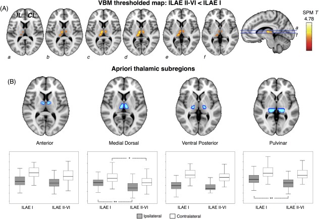
Voxel‐based morphometry (VBM) results: differences between patient outcome groups. (A) The single cluster of voxels (yellow–orange) significantly different when patient outcome groups were directly compared, which corresponded to regional thalamic atrophy in patients with persistent postoperative seizures. (B) Quantification of mean gray matter volume within 4 thalamic a priori regions of interest revealed that ipsilateral medial dorsal and pulvinar, and contralateral medial dorsal regions were significantly atrophic in patients with persistent postoperative seizures relative to those rendered seizure free. CL, contralateral; IL, ipsilateral; ILAE = International League Against Epilepsy; SPM = Statistical Parametric Mapping. *p < 0.05, **p < 0.01. [Color figure can be viewed in the online issue, which is available at www.annalsofneurology.org.]