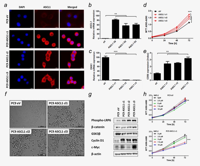
Figure 2.

ASCL1 expression induced “small cell‐ness” and canonical WNT‐signaling. (a) IF of PC9 transfected with ASCL1 expression plasmid (ASCL1) or empty vector (eV). Bars indicate 20 μm. (b+c) ASCL1 and DKK1 mRNA expression determined using qRT‐PCR. (d) Cell proliferation measured by MTT assay. (e) CD56 expression determined by flow cytometry. Mean fluorescence intensity of CD56‐PE‐Cy7 was normalized on IgG‐PE‐Cy7 (Index). (f) Cell morphologies determined in monolayer cell culture by microscopy. Bars indicate 100 μm. (g) Protein levels of members of WNT‐signaling pathway determined by Western blot. (h) eV transfected PC9 and an ASCL1 expressing representative clone c1 were treated with IWP‐2 WNT‐pathway inhibitor. Cell proliferation measured by MTT assay. Analysis was done using ΔΔCT‐method. Data are presented as mean ± SEM (n = 5). Statistical significance was calculated using a Student's t test, two‐sided, * p < 0.05, ** p < 0.01, *** p < 0.001.
