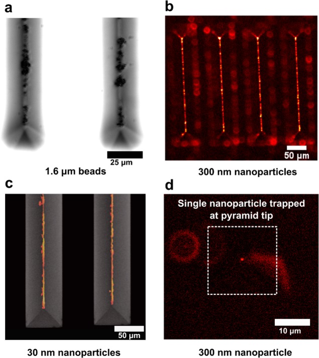
Figure 3.

Tip-enhanced trapping of magnetic nanoparticles. (a) Bright-field light microscopy image showing 1.6 μm beads captured on tips of wedges. (b) Fluorescence images showing 300 nm nanoparticles captured on sharp wedge tips. (c) Fluorescence image showing 30 nm magnetic nanoparticles captured on sharp wedge tips. The image has been overlaid on top of a SEM of the wedges. (d) Fluorescence image showing capture of a single 300 nm magnetic nanoparticle at the tip of one such pyramid under the influence of a magnetic field.
