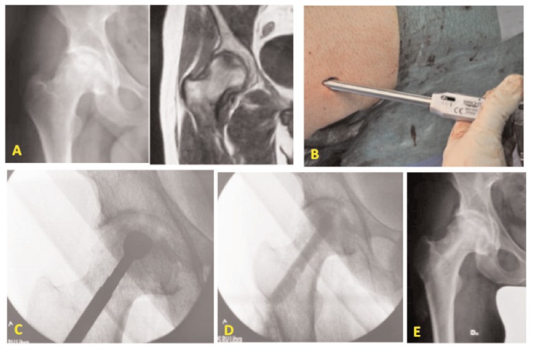
Figure 1.
Male patient of 44 years old with osteonecrosis of the right femoral head. A) Preoperative X-ray and MRI of the right hip; B) mini-invasive approach to the greater trochanter for the standard Core Decompression; C) burring the necrotic lesion in the femoral head through the femoral neck; D) fluoroscopic control at the end of the surgical procedure: conventional CD and application of bone substitute and autologous MSCs from iliac crest; E) X-rays at 3 months after surgical procedure.

