
Figure 1. Accumulation of γδ T cells in breast cancer but not in normal breast tissues.
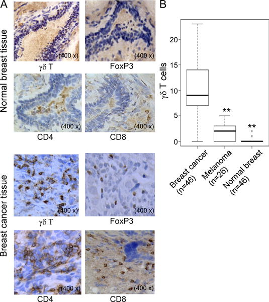
(A) Immunohistochemical staining of γδ, CD4+ and CD8+ T cells, as well as FoxP3+ cells in normal breast and cancer tissues. Few γδ, CD4+ and CD8+ T cells, as well as FoxP3+ cells were observed in normal breast tissues. However, high numbers of γδ, CD4+ and CD8+ T cells, as well as FoxP3+ cells were detected in breast cancer tissues. Frozen or paraffin-embedded tissue sections were immunohistochemically stained to detect the indicated cells. (B) Significantly increased numbers of γδ T cells existed in breast cancer tissues compared with normal breast tissues and melanoma tumor tissues. Frozen sections from breast tumor samples and controls of paired normal breast tissues (n=46) and melanoma tissues (n=26) were immunohistochemically stained to detect γδ T cells. Number of γδ T cells shown is the average numbers per high field (400 ×) in each tissue sample. The median number of γδ T cells in each group is shown as a horizontal line. Significance was determined by paired (breast cancer vs normal breast tissues) or unpaired (breast cancer vs melanoma tissues) T test. **p< 0.01, compared with γδ T cells in the breast cancer tissues.