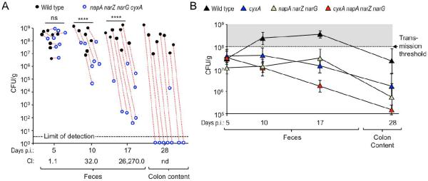
Figure 5. Cytochrome bd-II oxidase and nitrate reductases contribute to a luminal S. Typhimurium expansion in the absence of antibiotic treatment.
(A) Groups of CBA mice were infected intragastrically with a 1:1 mixture of the S. Typhimurium wild type (black circles) and the respiration-deficient napA narZ narG cyxA mutant (blue circles) and samples collected at the indicated days after infection (days p.i.). Dotted red lines connect strains recovered from the same animal. CI, competitive index; ****, P < 0.001; ns, not statistically significantly different; nd, not determined. (B) Groups of CBA mice (N = 4) were infected intragastrically with 1 × 10⁸ CFU/animal of one of the indicated S. Typhimurium strains and samples collected at the indicated days after infection.

